
MCAT Planner
How the Catfish Transmissible Cancer Explains MCAT Biology
A brown bullhead catfish with melanoma sounds like the kind of odd animal fact you can safely ignore during MCAT prep. It is not on the high-yield content list as “catfish cancer.” But the biology research behind the 2026 discovery is exactly the kind of unfamiliar passage that can punish a student who memorized vocabulary without practicing interpretation: tumor cells, host genetics, immune recognition, phylogeny, sequencing controls, and one central question hiding underneath the story.
In July 2026, a Nature paper reported that melanoma in brown bullhead catfish from Lake Memphremagog represents a naturally occurring transmissible cancer — the fourth known type, and the first described in fish and in a freshwater system.[1] The useful MCAT question is not “Can fish get contagious cancer?” It is: how did researchers show that tumors in different fish came from the same cancer lineage, instead of separate cancers, a shared environmental exposure, or an infectious agent causing new tumors in each host?
The claim only matters because the evidence is genomic
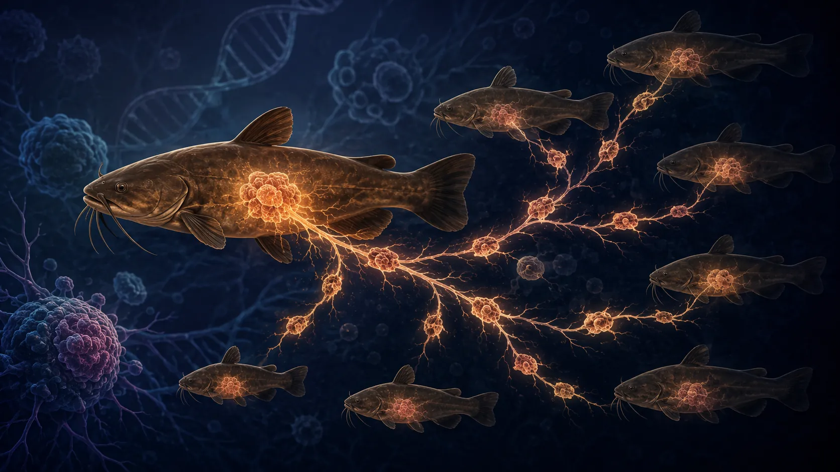
The word “transmissible” does a lot of work here. In ordinary cancer, a host’s own cells acquire mutations, expand clonally, and form a tumor. In a transmissible cancer, the cancer cell lineage itself moves between hosts and survives as a living graft. That means the tumor in one animal should carry genetic evidence of ancestry with tumors in other animals, not just evidence that each animal developed a similar disease.
The Nature study approached that problem with paired tumor-normal whole-genome sequencing from 19 fish.[1] That pairing is the part students should notice first. A tumor-only sample can tell you what mutations are present in the tumor. A matched normal sample helps identify which variants belong to the fish’s inherited genome and which belong to the tumor lineage. If the tumor really came from the host, its background genome should mostly match that fish. If the tumor was transmitted from another individual, the tumor genome should look partly foreign to the host that carries it.
The tumors shared single-nucleotide variants across different fish, a pattern that is hard to explain if each fish independently developed its own melanoma.[1] For an MCAT-style passage, this is clonal evolution in a clean form: descendants of one ancestral cancer cell inherit a recognizable set of mutations. Those inherited variants become a lineage marker. The tumor is not merely “similar”; it carries shared genomic scars.
The study also compared the catfish findings with 463 human melanomas from The Cancer Genome Atlas. In that comparison, 99.9% of human melanoma SNVs were unique to each patient.[1] That number is useful because it gives the contrast students often need: ordinary cancers are genetically personal to the patient, while a transmissible cancer should show a shared clonal signature across hosts.
| What the researchers compared | What the pattern means for interpretation |
|---|---|
| Tumor DNA vs. matched normal DNA from the same fish | Separates inherited host variants from tumor-lineage variants |
| Tumors from different fish | Tests whether tumors share a common clonal origin |
| Catfish tumors vs. human TCGA melanomas | Shows how unusual cross-host shared SNVs are compared with ordinary melanoma |
| Tumor sequence data vs. microbial and viral signals | Tests whether an infectious agent, rather than a cancer cell lineage, explains the disease |
The strongest passage clue is not one mutation; it is a pattern across methods
Shared mutations alone would be suggestive, but the stronger case asks whether multiple independent lines of evidence point in the same direction. That is where the catfish paper becomes more than a strange wildlife report.
Mitochondrial data supported the same interpretation. Tumor mitochondrial genomes formed a monophyletic clade distinct from the host fish, and the tumors showed mitochondrial heteroplasmy at 32–37 sites compared with 0–4 sites in normal tissue.[1] In passage terms, the tumor mitochondria were not behaving like a random sample of each host’s own mitochondrial background. They carried evidence of a shared tumor lineage.
Copy-number variation added another layer. Tumor-tumor CNV profiles were strongly correlated, with r = 0.83, while tumor-normal comparisons were much less correlated, with r = 0.22.[1] That matters because CNVs are large-scale genomic changes: duplications, deletions, and other dosage changes. When multiple tumors across different fish show similar CNV architecture, the simplest interpretation shifts toward shared ancestry rather than independent parallel cancers.
The authors also used Kraken2 metagenomic screening and DESeq2 analysis to test for viral or microbial explanations.[1] This is the part that many short science-news summaries flatten. A transmissible cancer is not the same thing as a virus causing cancer. If a virus caused separate melanomas in many fish, you might see a disease cluster without the tumor cells themselves being transmitted. The paper’s screening was aimed at ruling out that kind of alternative explanation.
So the argument is cumulative: paired tumor-normal sequencing, shared tumor SNVs, tumor mitochondrial phylogeny, mitochondrial heteroplasmy, CNV correlation, and metagenomic screening all push away from “many fish independently developed melanoma” and toward “one cancer lineage moved between hosts.”[1]
What the Lake Memphremagog timeline does — and does not — prove
The tumors were first noticed during surveys between 2012 and 2017, and the Nature paper reported prevalence values from 23% to 37% in 2014–2017 surveys.[1] Nature News framed the finding for a broader audience as a rare contagious cancer discovered in wild catfish, but the primary paper is where the genomic confirmation sits.[2]
That distinction matters for students. Observation of many affected fish can raise suspicion. It cannot, by itself, prove transmission of cancer cells. A shared environment could increase cancer risk. A pathogen could cause tumors. Genetic relatedness among fish could make similar cancers more likely. The paper’s sequencing design is what moved the claim from “unusual cluster” to “transmissible cancer lineage.”
There is also a press-release detail worth handling carefully. The University of Vermont highlighted historical context, including Henry David Thoreau’s 1852 observation of tumor-bearing bullheads, and discussed elevated arsenic in the setting.[3] Those details are interesting, but they are not the main proof of transmissibility. Elevated arsenic is a correlation in this context, not a demonstrated cause of the transmissible melanoma. If this were a passage, overclaiming that arsenic caused the cancer would be exactly the kind of tempting answer choice to avoid.
Why immune evasion becomes the unavoidable biology
A transmissible cancer has to solve a problem that ordinary cancer does not. It is not only evading immune surveillance inside the original host; it must survive after entering another genetically distinct host. That turns the case into a histocompatibility problem.
For MCAT purposes, this is where MHC should light up. MHC molecules present peptide fragments to T cells. If a grafted cell enters a new host, differences in histocompatibility molecules can mark it as foreign. A transmissible cancer therefore needs some way to persist despite immune recognition. The catfish paper identifies the transmissible lineage, but the exact transmission route and full immune-evasion mechanism remain unresolved.[1]
That uncertainty is not a flaw in the lesson. It is the lesson. A passage may give you strong evidence for clonal transmission while leaving the mechanism of spread as a hypothesis. In catfish, possibilities such as spawning contact or waterborne transmission are under investigation, not settled facts.[1]
The other transmissible cancers make the immune logic easier to see
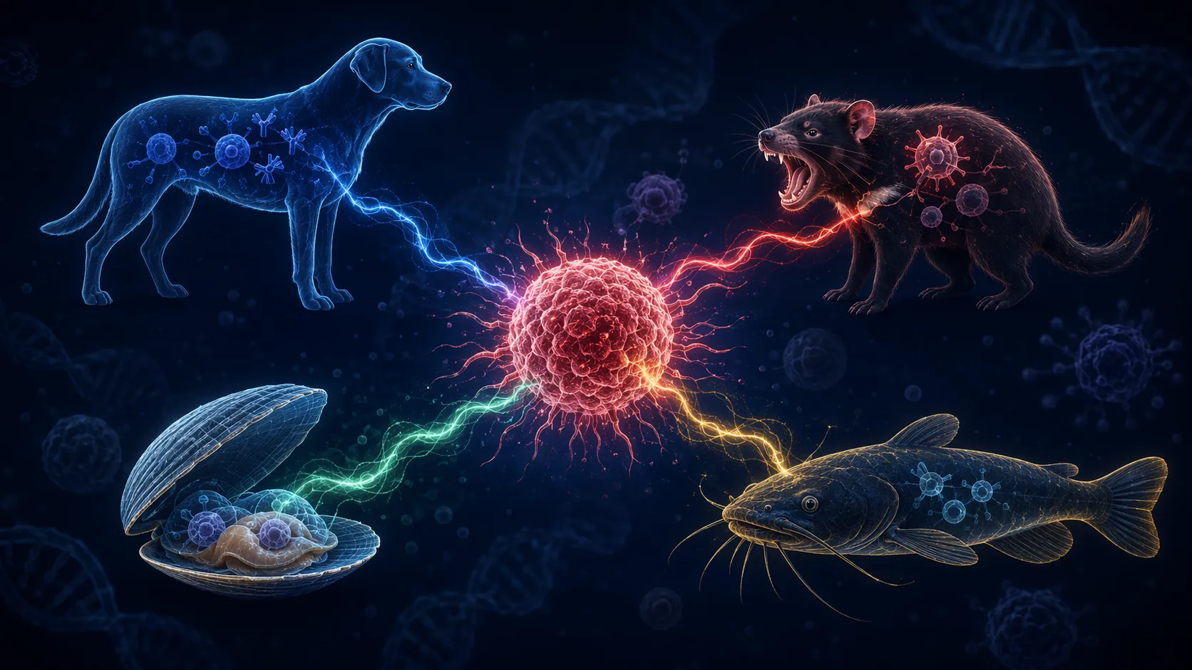
The catfish melanoma now sits beside three previously known naturally occurring transmissible cancer systems: canine transmissible venereal tumor in dogs, devil facial tumor disease and DFT2 in Tasmanian devils, and bivalve transmissible neoplasia.[4][5] The comparison is not just trivia. Each system forces the same question: how can a cancer cell lineage persist after moving into another body?
| Transmissible cancer | Host group | MCAT concept it helps practice |
|---|---|---|
| Canine transmissible venereal tumor | Dogs | Tumor as a long-lived clonal cell lineage; immune evasion and graft-like survival |
| Devil facial tumor disease / DFT2 | Tasmanian devils | Histocompatibility barriers, MHC-I expression, and immune recognition |
| Bivalve transmissible neoplasia | Bivalves | Transmission without vertebrate-style MHC constraints |
| Brown bullhead catfish melanoma | Freshwater fish | Tumor-normal genomics, clonal origin, phylogeny, and unresolved transmission mechanism |
Canine transmissible venereal tumor is often described as the oldest known continuously living mammalian cancer cell lineage, with estimates around 11,000 years.[5] That should sound biologically strange: the cancer is not merely recurring in dogs as a disease category; the cell lineage itself has persisted across many hosts over evolutionary time.
Tasmanian devil facial tumor disease gives the clearest immunology hook. Devil facial tumor cells have been associated with MHC-I downregulation, helping tumor cells avoid normal immune detection.[5] If a passage asks why reduced MHC-I expression could help a transmissible tumor, the answer is not that MHC-I is “bad.” It is that antigen presentation can expose abnormal or foreign cells to cytotoxic T-cell recognition. Reducing that display can help the tumor hide.
Other immune-evasion routes can matter too. TGF-β-mediated immune suppression is one mechanism discussed in transmissible cancer biology, and it maps neatly onto the broader cancer hallmark of avoiding immune destruction.[5] The bivalve case is different because bivalves lack the vertebrate MHC system, so the histocompatibility barrier is not the same barrier a mammal or fish tumor must navigate.[5]
For a side-by-side comparison of all four transmissible cancers, see our companion reference on the first freshwater transmissible cancer. The important study habit is to compare them along one dimension at a time: host, transmission evidence, immune barrier, and mechanism known versus unknown.
How this becomes an MCAT passage instead of a headline
No one needs to add “brown bullhead melanoma facts” to a flashcard deck as if the AAMC announced a new organism of the month. The relevance is inferred from AAMC content categories — cancer biology, immune recognition, genetics, evolution, and experimental interpretation — and from the precedent of transmissible cancers such as DFTD and CTVT appearing in practice-style materials. That is different from claiming that the catfish paper itself is now an official MCAT topic.
What is worth practicing is the reasoning move. An MCAT passage could describe an unfamiliar organism, give sequencing data, mention host and tumor samples, and ask what conclusion is best supported. The strongest students will not panic over the animal. They will translate the story back into core biology.
- If tumors from different hosts share many variants, ask whether that supports a common clonal origin.
- If tumor DNA differs from matched normal DNA, ask whether the tumor may have a distinct lineage from the host.
- If a microbial or viral screen is negative, do not describe the disease as pathogen-caused without additional evidence.
- If an environmental contaminant is elevated, separate correlation from causation.
- If a cancer moves between hosts, expect immune evasion or an unusual histocompatibility context to be part of the explanation.
This is also a good case for practicing how to read primary research without drowning in it. If that is the skill you are building, the same research-reading approach appears in our guide on using Greenland shark studies to read scientific research. The animal changes. The passage behavior does not.
A compact way to study the catfish case
When reviewing this discovery, do not start by memorizing the lake, the fish, or the headline label. Start with the hypothesis tree:
- Separate cancers in separate fish: would predict mostly host-specific tumor genomes.
- A shared carcinogenic environment: could explain increased cancer occurrence, but not by itself shared tumor lineage markers.
- A virus or microbe causing tumors: would require evidence of an infectious agent driving new cancers.
- A transmitted tumor clone: predicts shared tumor SNVs, shared mitochondrial lineage patterns, and correlated tumor CNVs across hosts.
The Nature paper’s evidence fits the fourth model best.[1] That is the answer path a passage would reward: not recognition of a rare fact, but elimination of weaker explanations using the data given.
If you are building a broader MCAT review plan, this catfish example pairs well with other real-science MCAT applications: the 2026 Cyclospora outbreak case study, the article on an engineered probiotic and pancreatic cancer, and the MCAT study prep hub for organizing content review around passage interpretation.
The practical judgment is simple: the catfish transmissible melanoma matters for MCAT prep not because students must memorize brown bullhead biology, but because it gives a current, concrete model for turning unfamiliar research into tested biology — clonal evolution, immune evasion, histocompatibility, and genomic evidence all in one passage-ready case.
References
- Brown bullhead catfish melanoma represents a novel transmissible cancer, Nature, July 22, 2026
- Rare contagious cancer discovered in wild catfish, Nature News
- More Like Parasites Than Tumors, University of Vermont
- Scientists Just Discovered A Contagious Cancer In A Catfish, The Fourth Known To Science. Meet The Other Three, Forbes
- Transmissible Cancers in an Evolutionary Perspective, PMC, 2020
Fill in this timeline
This is a skeleton schedule, not a performance claim — for section-by-section strategy to fill in each slot, read the exam hub. For evidence that a similar timeline worked, compare against real outcome logs.
How to Read the '1 in 4 NFL Players CTE' Study
- When to Do HIIT for Better Memory and Exam Focus
- Can NYT Strands Daily Actually Build Vocabulary?
- A Sleep-First Routine to Stop Falling Asleep While Studying
- Does Air Quality Affect Study Focus? Yes — by How Much?
Compare each outcome's duration against this planner's 8 weeks length manually.