
MCAT Exam Hub
Learn MCAT Parasitology from the 2026 Cyclospora Outbreak
Use the 2026 multistate Cyclospora outbreak — the largest documented in US history — as a single real-world case study to master apicomplexan biology, the complete life cycle, the critical distinction from Cryptosporidium (which explains why no direct transmission occurs), the diagnostic methods tested on the MCAT, and the TMP-SMX treatment regimen.
- gre
- mcat
- asvab
- sat
- act
- digital-adaptive
- official-material
- section-strategy
- test-date-timeline
A useful Cyclospora outbreak case study for medical students starts like a passage stem, not a headline: in July 2026, CDC reported a multistate cyclosporiasis outbreak linked to iceberg lettuce from Taylor Farms de Mexico served at Taco Bell. As of the July 20 investigation update, CDC listed 4,173 laboratory-confirmed cases, 308 hospitalizations, no deaths, and affected patients in 41 states; 90% of interviewed patients reported eating iceberg lettuce before illness. CDC described it as the largest documented Cyclospora outbreak in U.S. history, but those figures are timestamped because the investigation was ongoing in Q3 2026.[1]

That is enough outbreak detail to establish the case. The exam question begins one layer deeper: what must be true about this parasite for thousands of produce-associated infections to occur without direct person-to-person spread?
The Organism Hiding Inside the Lettuce Stem
Cyclospora cayetanensis is a coccidian protozoan in the phylum Apicomplexa. That classification is not decorative. Apicomplexans are intracellular parasites, and Cyclospora’s clinically important site is the small intestine, where infection produces watery diarrhea that can be prolonged or relapsing if untreated.[2]
For MCAT and Step-style reasoning, the organism’s label should immediately pull three ideas into view: protozoan parasite, intestinal epithelial infection, and oocyst-based transmission. The 2026 outbreak then supplies the real-world wrapper: fresh produce can carry the infective form, many people can be exposed through a shared ingredient, and routine foodborne-bacteria reflexes will not explain the biology.
| Case-stem clue | What it should make you think |
|---|---|
| Prolonged watery diarrhea after fresh produce | Intestinal protozoan; Cyclospora belongs high on the list |
| Large multistate cluster linked to lettuce | Common-source foodborne exposure, not household spread |
| No direct person-to-person transmission expected | Oocysts are not infective immediately after being shed |
| Routine O&P may be negative | Special stains, autofluorescence, or PCR are needed |
| Treatment response to TMP-SMX | Classic cyclosporiasis therapy |
The Life Cycle Is the Answer, Not a Diagram to Memorize
Start at the moment of exposure. A person ingests sporulated Cyclospora oocysts on contaminated food or water. “Sporulated” is the key word: this is the mature, infective form. After ingestion, the oocyst excysts in the gastrointestinal tract, releasing sporozoites that invade epithelial cells of the small intestine.[2]
Inside those intestinal cells, Cyclospora undergoes asexual multiplication by merogony. That gives the organism a way to amplify within the host before shifting into sexual development, or gametogony. Fertilization produces oocysts, which are then shed in stool.[2]
Here is the hinge that explains the outbreak pattern: the oocysts shed in stool are unsporulated. They are not immediately infective. CDC DPDx describes Cyclospora oocysts as requiring environmental sporulation, typically 7–14 days at 22–32°C, before they become infective.[2]
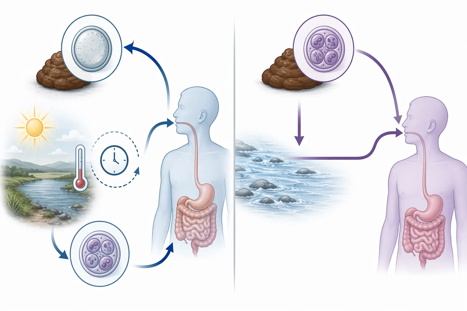
That one delay is why “infected patient has diarrhea” does not equal “infected patient directly infects the next person.” Freshly passed Cyclospora oocysts need time and environmental conditions before they can infect another host. A contaminated field, water source, packing environment, or produce-handling chain can therefore matter much more than bedside proximity.
This is also the cleanest place to compare Cryptosporidium. Cryptosporidium oocysts are shed already sporulated and infective, which makes direct fecal-oral transmission possible. Cyclospora oocysts are shed unsporulated and need environmental maturation, which is why direct person-to-person spread is not expected.[2][3]
If a test item gives two nearly identical protozoa and asks which one spreads directly in a daycare, hospital room, or household, the life-cycle state of the shed oocyst is doing the work. Cryptosporidium can fit that direct-transmission pattern. Cyclospora generally cannot, because the host sheds the wrong developmental stage.
The Complete Cyclospora Flow
- A person ingests sporulated oocysts from contaminated food or water.
- Excystation releases sporozoites in the gastrointestinal tract.
- Sporozoites invade small-intestinal epithelial cells.
- Asexual merogony expands the infection within enterocytes.
- Sexual gametogony produces oocysts.
- Unsporulated oocysts are shed in stool.
- Oocysts sporulate in the environment over 7–14 days at suitable temperatures, becoming infective for the next host.[2]
That sequence is more durable than a flashcard. It explains the foodborne source, the lack of direct spread, the stool-based diagnosis, and why an outbreak can look like a public-health mystery even though the clinical syndrome is just watery diarrhea after produce.
Why Routine O&P Can Miss the Diagnosis
The diagnostic trap is almost unfair in exactly the way exams like. A clinician suspects an intestinal parasite and orders an ova-and-parasite examination. The result can still miss Cyclospora because standard stool stains used in routine O&P workflows, including trichrome and iron hematoxylin, do not reliably detect Cyclospora oocysts.[4]
So the correct next move is not “parasites ruled out.” It is “ask whether the lab used a method that can actually see this organism.” Cyclospora oocysts can be detected with modified acid-fast staining, modified safranin staining, or ultraviolet fluorescence microscopy; CDC DPDx notes autofluorescence under ultraviolet excitation at 330–380 nm.[2]
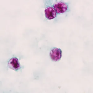
Modified acid-fast stain is the classic exam association. Cyclospora oocysts are round and variably acid-fast, so they may stain pink to red while others stain faintly or not at all. That variability is not a reason to discard the diagnosis; it is part of the organism’s diagnostic profile.[2]
Autofluorescence is a second high-yield clue because it turns the organism’s physical properties into a lab answer. Under the right ultraviolet excitation, the oocysts fluoresce, allowing detection when routine staining has failed. Safranin staining is another specialized approach, less likely to be the first association a student recalls but still part of the real diagnostic toolkit.[2]
Multiplex PCR is the newer layer. CDC’s clinical overview notes that molecular diagnostic tests, including multiplex gastrointestinal panels, can detect Cyclospora; the BioFire FilmArray GI Panel is a 22-target multiplex PCR panel and was first shown to detect Cyclospora during a 2018 Wisconsin outbreak.[4]
| Diagnostic method | How to treat it for exams |
|---|---|
| Routine O&P with standard trichrome or iron hematoxylin | Can miss Cyclospora; a negative result does not exclude it |
| Modified acid-fast stain | Classic tested method; oocysts may stain variably acid-fast |
| Modified safranin stain | Specialized stool stain that can detect oocysts |
| UV autofluorescence | High-yield visual clue; oocysts autofluoresce at specified UV wavelengths |
| Multiplex PCR | Modern clinical and outbreak tool; useful when panels include Cyclospora |
The student-level mistake is to memorize “Cyclospora = acid-fast” without understanding why the ordinary stool workup failed first. The clinical reasoning chain is the tested part: prolonged watery diarrhea after produce, routine O&P unrevealing, then special staining, autofluorescence, or PCR.
Treatment Is Short, Specific, and Testable
The treatment association is trimethoprim-sulfamethoxazole. CDC lists the typical adult regimen as one double-strength TMP-SMX tablet twice daily for 7–10 days.[4]
The efficacy claim is not just inherited board-prep folklore. In a placebo-controlled trial in Nepal published in 1995, 6.3% of treated patients still had detectable oocysts at day 7, compared with 88.2% of placebo recipients; that result supports the commonly cited greater-than-90% effectiveness of TMP-SMX for cyclosporiasis.[4]
For exams, the answer is usually not metronidazole, nitazoxanide by reflex, or supportive care alone. If the stem has Cyclospora’s biology and diagnostic clues, TMP-SMX should feel like the endpoint of the reasoning chain, not a detached drug fact.
What the 2026 Investigation Adds Without Taking Over
CDC’s 2026 investigation did not stop at “people ate at Taco Bell.” The July update described ingredient-level analysis using multivariable backward stepwise logistic regression, with iceberg lettuce emerging as the implicated ingredient and traceback pointing to Taylor Farms de Mexico.[1]
That kind of ingredient-level work is useful for research-passage literacy. It shows how public-health investigators move from a restaurant exposure to a specific food item, then to a supplier. It is not the center of the MCAT biology lesson, but it explains why a parasite life cycle can become a supply-chain investigation.
A smaller 2023 Alabama restaurant outbreak makes the same point with cilantro. In an MMWR report published in April 2025, investigators used a three-step analysis moving from menu items to ingredients to multivariable modeling; cilantro was associated with illness with an odds ratio of 40.9, but the 95% confidence interval was wide, 6.4–808.6, reflecting the limits of a small outbreak analysis.[5]
That wide interval matters. It does not erase the cilantro signal, especially alongside traceback findings, but it should keep a student from treating a dramatic odds ratio as if it were a precise measurement. On an exam passage, the statistical limitation may be the point.
Genotyping Is Beyond the Core Card, but Good Passage Material
CDC’s CYCLONE system is one of those details that is usually beyond basic MCAT recall but very fair game for interpreting a research passage. CDC describes CYCLONE as an 8-marker genotyping system, using 6 nuclear and 2 mitochondrial markers with deep sequencing, and reports 93.8% sensitivity and 99.7% specificity for outbreak cluster identification.[6]
The important distinction is adoption versus biological explanation. Genotyping can help decide whether clinical specimens cluster together during an outbreak. It does not replace the life-cycle logic that explains why the organism arrives through contaminated produce rather than direct contact.
Cyclospora research also has a practical constraint that would fit neatly into an experimental-design question: C. cayetanensis cannot currently be propagated in routine in vitro culture or in an animal model, so investigators rely heavily on clinical specimens, environmental detection, and molecular methods.[7]
There is also preliminary molecular evidence that what has been called C. cayetanensis may include more than one human-associated species, including proposed C. ashfordi and C. henanensis. That is useful context for advanced readers, but it has not replaced the clinical and exam-level organism name most students are expected to know.[7]
The Exam Takeaway
If a passage gives prolonged watery diarrhea after fresh produce, a common-source outbreak, no expected direct person-to-person spread, a negative or unrevealing routine O&P, oocysts detected by modified acid-fast stain, safranin stain, UV autofluorescence, or multiplex PCR, and response to TMP-SMX, the organism should reconstruct itself.
Cyclospora is not just “the lettuce parasite” or “the TMP-SMX parasite.” It is an apicomplexan coccidian protozoan whose unsporulated shed oocysts must mature in the environment before becoming infective. Once that mechanism is clear, the outbreak pattern, the Cryptosporidium contrast, the diagnostic trap, and the treatment choice line up in one case.
References
- Investigation Update: Cyclospora Outbreak Linked to Iceberg Lettuce, CDC, July 2026,
- Cyclosporiasis, CDC DPDx,
- Cyclosporiasis: Updates on Clinical Presentation, Pathology, Clinical Diagnosis, and Treatment, PMC, 2021,
- Clinical Overview of Cyclosporiasis, CDC,
- Notes from the Field: Cyclosporiasis Outbreak Associated with a Restaurant — Alabama, 2023, MMWR, April 2025,
- Cyclosporiasis Surveillance, CDC,
- Cyclospora cayetanensis: Current Understanding of an Emerging Foodborne Parasite, PMC,
Related exhibits & inventory
Verified outcomes
Planners
No planner filed for this exam yet
A downloadable timeline template for this exam hasn't been published yet.
Tool verdicts
Questions about this plan
Ask a question about a specific section, timeline, or citation in this plan — or flag something that needs correcting.
Comments
Join the discussion with an anonymous comment.