
MCAT Exam Hub
MHC I Antigen Presentation and Arginine Study Notes for MCAT
Consolidated MCAT study notes for the MHC class I endogenous antigen-presentation pathway and arginine's basic-amino-acid chemistry, plus the July 2026 Cell finding that arginine availability controls codon-dependent MHC class I translation. The study is labeled frontier research for passage practice — not AAMC-tested content — so testable facts stay separate from the research headline.
- gre
- mcat
- asvab
- sat
- act
- digital-adaptive
- official-material
- section-strategy
- test-date-timeline
Keep three boxes separate before you make any cards: MHC class I antigen presentation is exam-ready immunology; arginine chemistry is exam-ready amino acid content; the July 2026 arginine–MHC I paper is frontier research that belongs in your passage-reasoning brain, not in the “AAMC definitely wants this mechanism memorized” pile.
| Topic | What to memorize | What to recognize in a passage |
|---|---|---|
| MHC class I | All nucleated cells; endogenous/cytosolic antigen; short peptide on MHC I; CD8+ cytotoxic T-cell recognition; proteasome → TAP → ER loading → Golgi → surface | How a virus, tumor cell, or experimental manipulation changes antigen display |
| Arginine | Basic amino acid; guanidinium side chain; positively charged at physiological pH; encoded by six codons | How amino acid availability, tRNA charging, codons, or translation speed could affect protein output |
| 2026 Cell arginine study | Do not memorize as settled MCAT content | Use as a mechanism passage: arginine restriction → arginyl tRNA repression → ribosome stalling at arginine codons → less MHC I translation → reduced antigen display |
If you only have ten minutes, start with the pathway. The test is much more likely to punish a scrambled MHC I sequence than to ask whether you remember a brand-new codon-dependence paper.
The MHC I pathway you actually need cold
MHC class I molecules are built to show the inside of a cell to CD8+ cytotoxic T cells. Structurally, classical MHC I is a heterodimer: a heavy chain of about 45,000 molecular weight paired with β2-microglobulin of about 12,000 molecular weight. The α1 and α2 domains of the heavy chain form the peptide-binding groove, and that groove binds short peptides, usually 8–10 amino acids long.[1]
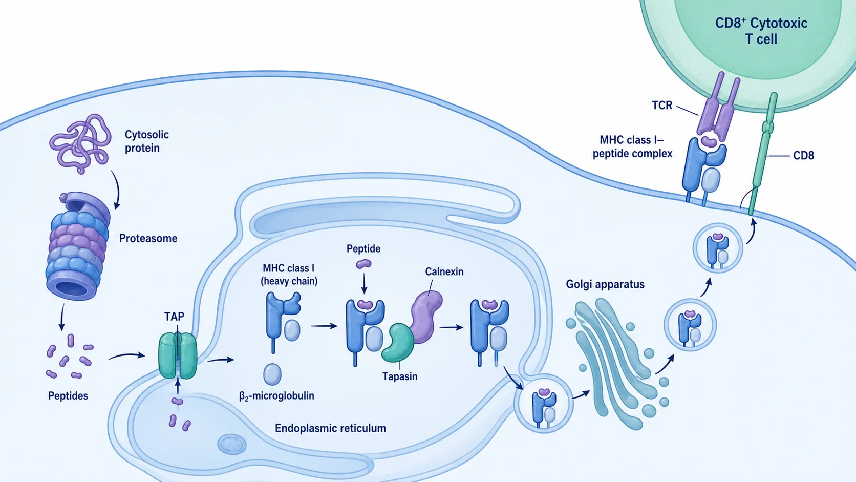
The clean sequence is:
- A cytosolic protein is degraded by the proteasome.
- The resulting peptides are transported into the endoplasmic reticulum by TAP.
- The MHC I heavy chain folds and assembles with β2-microglobulin with help from ER chaperones.
- A peptide is loaded into the MHC I groove, with tapasin helping connect the MHC I loading complex to TAP.
- The loaded MHC I–peptide complex moves through the Golgi.
- The complex reaches the plasma membrane and can be surveyed by CD8+ cytotoxic T cells.
That order is not decorative. Proteasome first, TAP second, ER loading third. If a passage knocks out TAP, peptides made in the cytosol do not efficiently enter the ER for loading onto MHC I. If β2-microglobulin is missing, stable surface expression of MHC I is disrupted. If peptide loading fails, the CD8+ T cell has less to inspect at the surface.
Where the peptides come from
For MCAT purposes, MHC I presents endogenous antigen: material generated inside the cell. That can include normal self proteins, viral proteins made inside an infected cell, or altered tumor-associated proteins. The proteasome cuts cytosolic proteins into peptide fragments that can feed the MHC I pathway.[1]
One useful detail for passage reasoning is DRiPs, or defective ribosomal products. These are newly synthesized proteins that are misfolded, prematurely terminated, or otherwise defective. A review of the MHC I pathway describes DRiPs as a major source of peptides for rapid sampling of newly synthesized proteins, which makes biological sense: an infected cell does not want to wait until every viral protein gets old before showing evidence of infection.[1]
Do not turn DRiPs into a separate memorization universe. The exam-useful idea is that MHC I samples intracellular protein production, and proteasomal degradation supplies peptides for that sampling.
TAP and ER loading
TAP is the transporter that moves peptides from the cytosol into the ER. That matters because MHC I folding and peptide loading happen in the ER, not out in the cytosol. Once inside the ER, peptides can be loaded onto the MHC I molecule as it assembles with help from several accessory proteins.[1]
The chaperone names are worth recognizing even if you do not make a giant card for each one: calnexin, calreticulin, ERP57, and tapasin. In the reviewed pathway, calnexin assists early folding of the MHC I heavy chain, calreticulin and ERP57 participate in the peptide-loading complex, and tapasin helps bridge MHC I loading with TAP so suitable peptides can be loaded efficiently.[1]
After a suitable peptide is loaded, the MHC I–peptide complex exits the ER, passes through the Golgi, and is delivered to the plasma membrane. At the surface, a CD8+ cytotoxic T cell can inspect the peptide. If the peptide signals infection or abnormal intracellular protein production in the right immune context, the CD8+ T cell can kill the presenting cell.
Do not mix up MHC I and MHC II
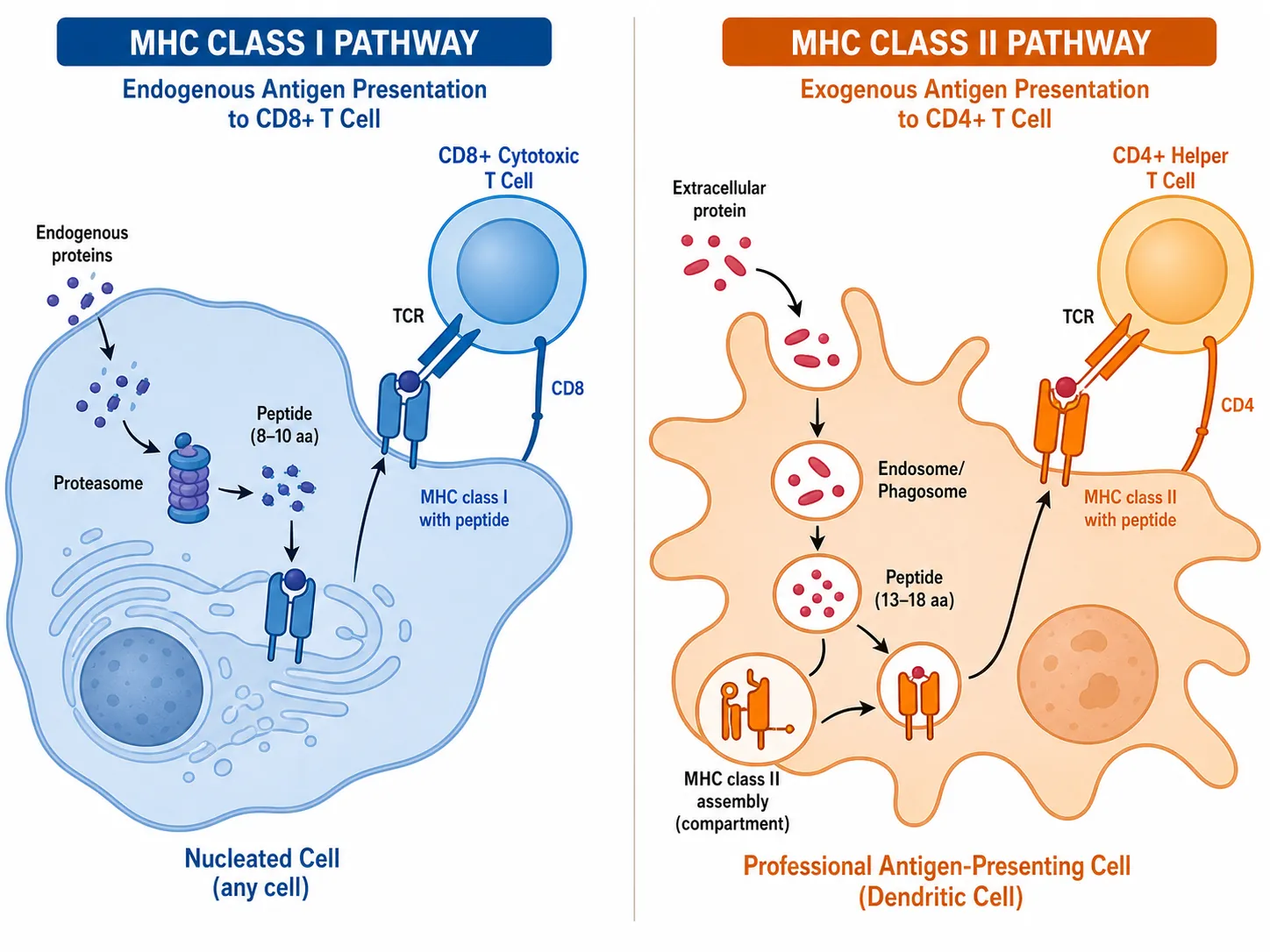
The classic exam trap is swapping the source of antigen or the T-cell partner. MHC I is broadly expressed on nucleated cells and presents endogenous antigen to CD8+ T cells. MHC II is restricted to professional antigen-presenting cells such as dendritic cells, macrophages, and B cells, and it presents exogenous antigen to CD4+ helper T cells. MCAT prep resources commonly frame the contrast this way.[2][3]
| Feature | MHC I | MHC II |
|---|---|---|
| Main expression pattern | All nucleated cells | Professional antigen-presenting cells: dendritic cells, macrophages, B cells |
| Antigen source | Endogenous/cytosolic | Exogenous/endocytosed |
| T-cell partner | CD8+ cytotoxic T cells | CD4+ helper T cells |
| Core exam association | Infected or abnormal cell shows what is happening inside | APC shows what it has taken up from outside |
For more immunology review in the same exam-first style, keep this near your broader MCAT hub notes rather than letting the topic drift into general immune-system news.
Arginine, first as an amino acid
Before arginine becomes part of an immunology headline, it is just one of your amino acids. Make that card simple.
- Arginine is a basic amino acid.
- Its side chain contains a guanidinium group.
- It is positively charged at physiological pH.
- It is encoded by six codons.
The first three bullets are standard amino-acid chemistry. The six-codon detail becomes unusually important in the 2026 paper because the mechanism depends on arginine codons and arginyl tRNAs, not just on “arginine is good for immunity.” A Rockefeller-linked report about the study also highlights arginine as a basic amino acid with a positively charged guanidinium group and notes that it is encoded by six codons.[4]
The July 2026 Cell paper: good passage material, not a new MCAT law
The new finding is real and interesting. In a Cell paper published online on July 30, 2026, Wu and colleagues reported that dietary arginine availability can control MHC class I translation in a codon-dependent way, with immune consequences in cell-culture and mouse models.[5][6]
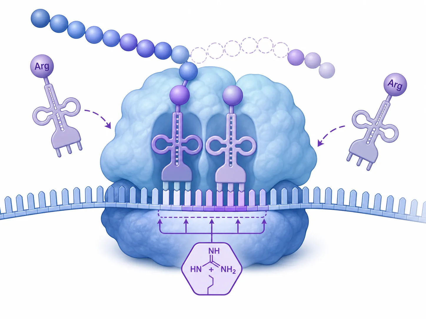
Here is the mechanism in passage language:
- Arginine availability falls.
- Arginyl tRNAs are repressed or become less available for efficient translation.
- Ribosomes stall at arginine codons on MHC class I transcripts.
- MHC class I translation drops.
- MHC I antigen display decreases.
- CD8+ T-cell recognition has less MHC I–peptide display to work with.
The elegant part is the codon-dependence. The authors reported that synonymous codon mutations prevented the effect, which supports the idea that the mechanism is tied to arginine codon usage during translation rather than simply to a broad collapse in immune signaling.[5][6]
That is exactly the kind of mechanism an MCAT-style passage could hand you. You would not need prior knowledge of the paper. You would need to track amino acid availability, charged tRNAs, codons, ribosome stalling, protein translation, and antigen presentation. In other words, it connects several testable topics without making the paper itself testable.
What the mouse and cell-culture results do — and do not — show
The reported immune outcomes are preclinical. In the Cell abstract and PubMed record, the authors report that dietary arginine restriction impaired antiviral immunity in influenza and SARS-CoV-2 mouse models and increased colon tumorigenesis in mice. They also report that supplementation or myeloid-specific deletion of arginase 1 had the opposite effect, and that these effects were abolished in β2-microglobulin-deficient mice.[5][6]
That β2-microglobulin detail is not random. β2-microglobulin is part of MHC I structure, so loss of the effect in β2-microglobulin-deficient mice supports the interpretation that the observed immune changes depended on the MHC I antigen-presentation axis rather than on some unrelated benefit of arginine.
The same boundary matters for supplementation. A secondary article in Inside Precision Medicine framed the work as relevant to how well T cells see tumors and viruses, but it also treated supplementation as a question for future trial design, not as proof that people should take arginine to improve cancer or viral outcomes.[7] The available reports do not give human trial results, dosing guidance, timing rules, or clinical benefit data.
So the safe study-note version is narrow: arginine scarcity can reduce MHC I translation through a codon-dependent mechanism in the reported experimental systems. Do not expand that into “arginine supplements improve immunity in humans.”
How this could appear in a B/B passage
A passage does not have to ask, “What did Wu et al. show in 2026?” It could give you the mechanism and then test whether you can reason from it.
- If arginine is restricted and MHC I translation falls, predict reduced MHC I surface display.
- If synonymous codons are changed so the transcript uses fewer vulnerable arginine codons, predict that the arginine-restriction effect may weaken.
- If TAP is knocked out, the issue is peptide transport into the ER, not codon-dependent translation.
- If β2-microglobulin is absent, surface MHC I stability becomes the problem.
- If a cell presents extracellular bacterial fragments to CD4+ T cells, you have probably left the MHC I pathway and entered MHC II territory.
Notice the sorting. Proteasome/TAP/ER loading questions are antigen-processing questions. Arginyl tRNA and ribosome-stalling questions are translation questions. CD8+ recognition questions are immune-effector questions. The passage may connect them, but your notes should not blur them.
If you like studying immunology through cases, pair this with Plague Vaccine as an MCAT Immunology Case Study or Use Pet Immunization Science to Master MCAT Biology Concepts. For the evidence-tiering problem — how far a dietary mouse study can travel before it becomes a human recommendation — the closer neighbor is How to Read Protein-Restriction Longevity Research.
What goes on the Anki card tonight
Put the stable material on cards:
- MHC I is expressed on all nucleated cells.
- MHC I presents endogenous/cytosolic peptides to CD8+ cytotoxic T cells.
- MHC I consists of a heavy chain plus β2-microglobulin.
- The MHC I α1/α2 groove binds short peptides, usually 8–10 amino acids.
- Endogenous pathway order: proteasome → TAP → ER loading with chaperones/tapasin → Golgi → surface display.
- MHC II presents exogenous antigen to CD4+ helper T cells and is found on professional APCs.
- Arginine is basic, has a guanidinium side chain, is positively charged at physiological pH, and is encoded by six codons.
Keep the 2026 Cell study as a reasoning example: arginine availability can affect arginyl tRNAs, ribosome movement at arginine codons, MHC I translation, and antigen display in the reported preclinical systems. Do not treat supplementation, clinical benefit, or AAMC-tested status as established.
References
- The MHC class I antigen presentation pathway: strategies for viral immune evasion — Immunology, 2003
- Major Histocompatibility Complex — Immune System — MCAT Content — Jack Westin
- Immune System: Antigen Presentation — MCAT Biology — MedLifeMastery
- A simple supplement could help the immune system fight cancer and viruses — ScienceDaily/Rockefeller, 2026
- Dietary arginine drives codon-dependent MHC class I translation and improves immunity in colon tumorigenesis and respiratory viral infection — Cell, July 30, 2026
- Dietary arginine drives codon-dependent MHC class I translation and improves immunity in colon tumorigenesis and respiratory viral infection — PubMed, 2026
- Arginine Availability May Shape How Well T Cells See Tumors and Viruses — Inside Precision Medicine
Related exhibits & inventory
Verified outcomes
Planners
No planner filed for this exam yet
A downloadable timeline template for this exam hasn't been published yet.
Tool verdicts
Questions about this plan
Ask a question about a specific section, timeline, or citation in this plan — or flag something that needs correcting.
Comments
Join the discussion with an anonymous comment.