
MCAT Exam Hub
Early Heart Disease Signs, Explained by Mechanism
Exam questions rarely reward a memorized list of heart disease symptoms — they reward connecting an early, subtle sign to the mechanism behind it. This med-school-level study guide organizes early signs by the four disease families exams test (ischemia, pump failure, arrhythmia, valve disease), pairs each sign with why it happens, and ends with a recall table, mnemonics, and a worked exam vignette.
- gre
- mcat
- asvab
- sat
- act
- digital-adaptive
- official-material
- section-strategy
- test-date-timeline
The phrase “early heart disease signs” looks friendly until an exam stem turns it into a trap. A student writes “fatigue, SOB, chest pain” in the margin, then misses the actual question because the stem is not asking for a symptom list. It is asking what physiology produced the clue.
Shortness of breath after climbing stairs can be an ischemic equivalent, a left-sided heart failure clue, a valve-flow problem, or an arrhythmia dropping cardiac output. Fatigue can mean underperfusion, reduced forward flow, or deconditioning from chronic congestion. Chest pressure can be stable angina; chest discomfort plus a separate acute-event pattern is a different testable moment. The same word does not keep the same mechanism.
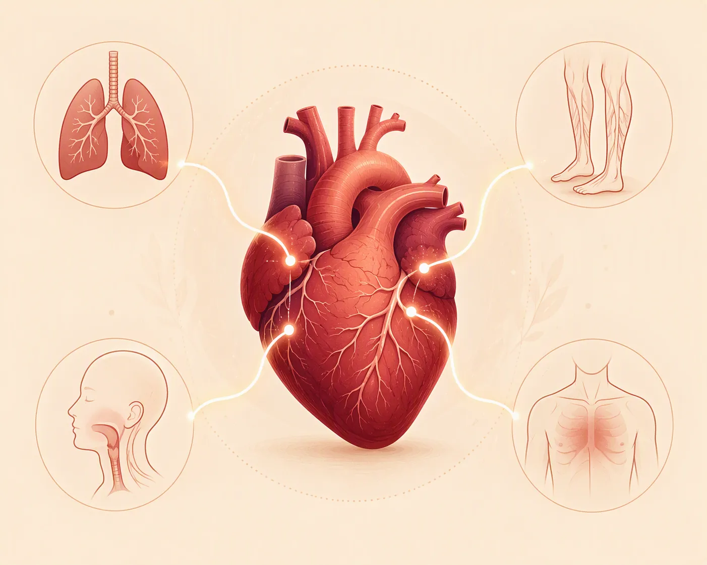
Use this as med-school-level support for vignette reasoning. It is useful for MCAT Bio/Biochem cardiovascular review, especially if you are building from a broader MCAT study plan, but it is not pretending to be a full USMLE course. The payoff is narrower and more testable: when a symptom appears early, ask whether the stem is pointing to underperfusion, pump failure, congestion, or conduction.
Start by splitting “heart disease” into exam families
CDC material treats heart disease as an umbrella, not a single diagnosis: it includes coronary artery disease, arrhythmias, cardiomyopathy, heart valve disease, congenital heart defects, and other conditions. That framing matters because early signs change when the failing part changes — vessel, myocardium, valve, or conduction system [1].
For exam purposes, the cleanest first pass is four families:
| Disease family | Early-sign mechanism to look for | Vignette translation |
|---|---|---|
| Coronary artery disease / ischemia | Supply-demand mismatch and myocardial underperfusion | Exertional pressure, dyspnea, or referred discomfort before an acute event |
| Heart failure | Reduced forward output plus backup of pressure into lungs or systemic veins | Fatigue, activity limitation, orthopnea, edema, rales, S3 |
| Arrhythmia | Abnormal conduction causing irregular rate, poor filling, or reduced output | Palpitations, episodic lightheadedness, near-syncope, exertional intolerance |
| Valve disease | Obstruction or regurgitation altering pressure and volume load | Murmur plus exertional dyspnea, fatigue, syncope, edema, or pulmonary congestion |

This is the same kind of pattern discipline used in other high-yield cardiology study tasks, such as separating lipid-risk categories in cholesterol guideline memorization or sorting statin indications in statin benefit groups for USMLE Step 1. The name of the symptom is only the first retrieval cue. The mechanism is what keeps the answer choices from blurring together.
Coronary artery disease and ischemia: early clues are often demand-related
The classic ischemia clue is not “pain” in the vague way students write it on a flashcard. Mayo Clinic describes angina as chest pain or discomfort that may feel like heaviness, pressure, squeezing, pain, burning, numbness, fullness, or a “tight band” around the chest. Mayo also notes that discomfort can refer to the neck, jaw, throat, upper belly, or back, and that shortness of breath can occur with coronary artery disease [2].
Mechanism first: ischemic myocardium is not receiving enough oxygenated blood for its current workload. Early on, the clue may appear when demand rises — walking uphill, carrying groceries, climbing stairs, emotional stress — then improve when demand falls. That is why “exertional” is doing so much work in a stem. It tells you the symptom is tied to supply-demand mismatch rather than being a random complaint floating in the history.
The early-vs-classic distinction becomes testable like this:
- Early or subtle ischemia: exertional chest pressure, exertional dyspnea, reduced exercise tolerance, or referred discomfort that resolves with rest.
- Classic presentation: angina descriptors such as pressure, squeezing, heaviness, or tight-band discomfort, especially with radiation to jaw, neck, throat, upper abdomen, or back.
- Exam disguise: the stem may avoid the word “pain” and instead say the patient feels chest heaviness, cannot finish the usual walk, or has discomfort in the jaw with exertion.
Do not over-clean this into “all ischemia announces itself early.” Mayo notes that many people are not diagnosed with coronary artery disease until they have chest pain, a heart attack, or another acute event [2]. AHA News also describes silent heart attacks as common and often overlooked, which means the absence of dramatic chest pain does not rule out clinically important ischemic disease [3]. CDC separates heart attack material as an event-type presentation rather than treating it as the same thing as chronic early signs [4].
That boundary condition is exactly why a single public-facing warning list is weak for exams. If the stem is chronic and exertional, reason through ischemia and demand. If the stem is an acute event, do not force it back into “early signs.” If the stem is silent or atypical, let risk factors, ECG findings, biomarkers, or downstream complications carry more of the logic.
Heart failure: fatigue and dyspnea become useful only after you locate the backup
Heart failure is where vague symptom memorization causes the most damage. “Fatigue” by itself is a weak clue. “Fatigue because forward cardiac output is no longer meeting tissue demand” is stronger. “Shortness of breath because left-sided pressures are backing into the pulmonary circulation” is stronger still.
Harvard Health’s early heart failure warning-sign framing emphasizes symptoms such as fatigue, reduced activity tolerance, congestion, edema, and shortness of breath [5]. HFSA’s FACES acronym is useful because it packages the cluster in a way students can retrieve under time pressure:
- F — Fatigue
- A — Activity limitation
- C — Congestion
- E — Edema
- S — Shortness of breath
The acronym is not the answer. It is the retrieval shelf. Once FACES comes up, split the clues into forward failure and backward congestion.
| Clue | Mechanism | How it may appear early | What it can precede |
|---|---|---|---|
| Fatigue | Reduced forward cardiac output | Patient stops normal exercise sooner or needs more rest | Overt exercise intolerance and poor perfusion signs |
| Activity limitation | Low reserve when demand rises | Stairs, hills, or routine errands become difficult | Marked functional limitation |
| Shortness of breath | Pulmonary venous congestion, especially in left-sided failure | Dyspnea first appears with exertion | Dyspnea at rest, pulmonary edema |
| Orthopnea | Recumbency increases venous return and pulmonary congestion | Needs extra pillows or avoids lying flat | Waking breathless, pulmonary edema pattern |
| Paroxysmal nocturnal dyspnea | Delayed nighttime pulmonary congestion | Sudden nighttime breathlessness | More advanced left-sided congestion |
| Peripheral edema | Systemic venous congestion | Ankle swelling late in the day | JVD, hepatosplenomegaly, more obvious right-sided failure |
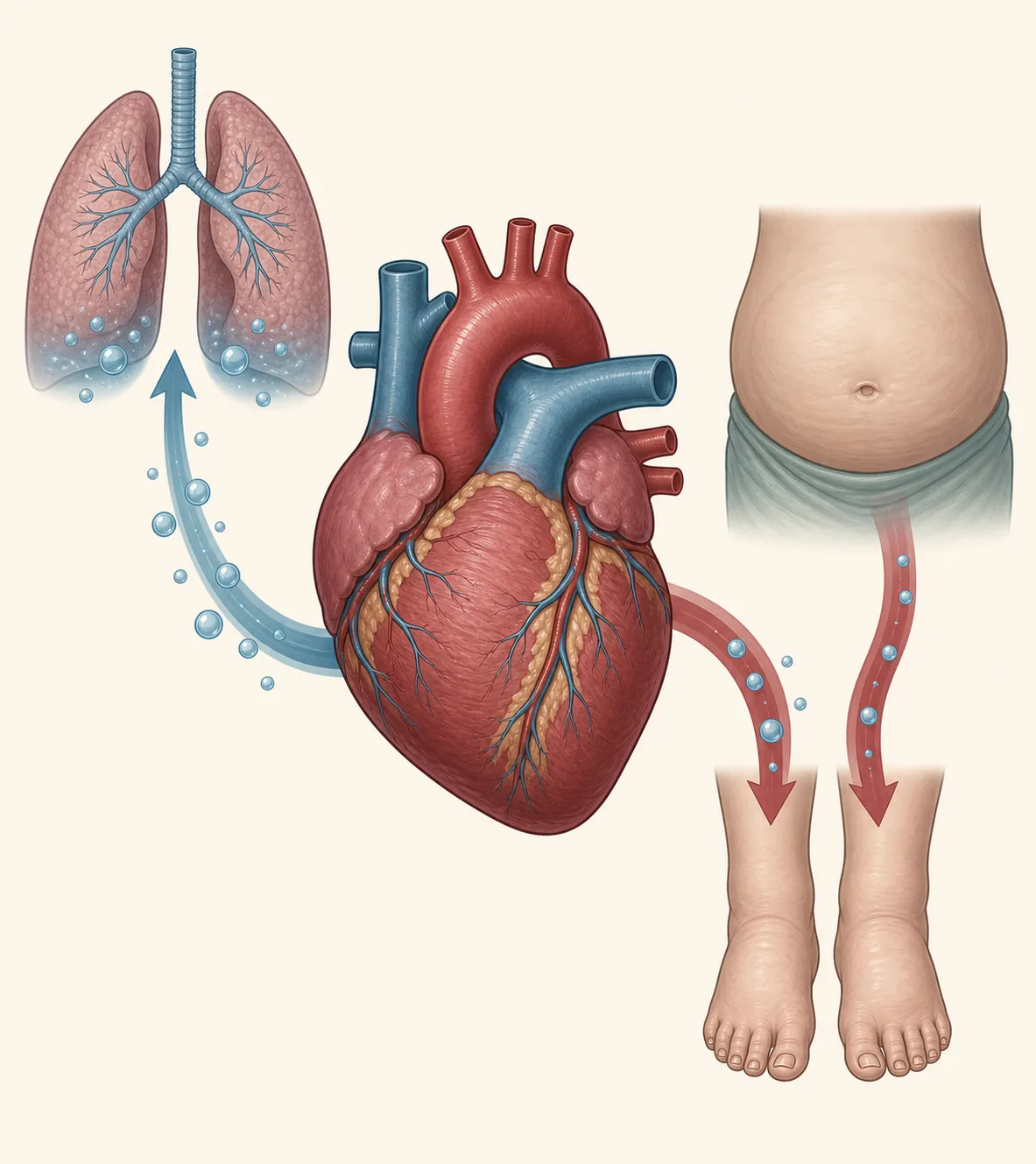
Osmosis’ left-heart-failure teaching vignette is useful here because it forces the left-vs-right split. Left-sided failure is associated with orthopnea, paroxysmal nocturnal dyspnea, and pulmonary edema, while right-sided failure is associated with jugular venous distention, hepatosplenomegaly, and peripheral edema. Osmosis also states the rule worth keeping in the front of your mind: left-sided heart failure is the most common cause of right-sided heart failure [6].
So when a stem says exertional dyspnea, rales, and an S3, do not stop at “shortness of breath.” Rales point toward fluid in the lungs. S3 points toward volume overload and a dilated, failing ventricle in the usual exam pattern. The mechanism is left-sided pump failure causing pulmonary venous congestion. If the patient later develops JVD and leg edema, that does not erase the left-sided process; it may show progression to right-sided involvement.
A common wrong move is to label all edema as “heart failure” and all dyspnea as “lungs.” In exam reasoning, edema must be located. Pulmonary congestion gives dyspnea, orthopnea, paroxysmal nocturnal dyspnea, rales, and pulmonary edema. Systemic venous congestion gives JVD, hepatosplenomegaly, and peripheral edema. Same broad disease family, different side of the circuit.
Arrhythmia: the early sign is often intermittent output failure
Arrhythmias belong in the heart disease umbrella, but their logic is electrical before it is congestive. The early clue is often episodic: a racing or irregular heartbeat, brief lightheadedness, near-syncope, exertional intolerance, or shortness of breath during episodes. Mayo’s heart disease symptom material separates irregular heartbeat symptoms from coronary and valve symptom patterns, and the British Heart Foundation also includes palpitation-type symptoms among signs that may suggest heart disease [2][7].
Mechanism first: abnormal rhythm can reduce stroke volume by making the rate too fast, too slow, or irregular enough to impair filling. If ventricular filling falls, cardiac output falls. The patient may feel palpitations, but the exam question may care more about what the palpitations do to perfusion: dizziness, presyncope, syncope, chest discomfort, or dyspnea.
| Stem clue | Mechanism move | What not to do |
|---|---|---|
| “Heart racing” with lightheadedness | Think tachyarrhythmia reducing filling time and output | Do not file it only under anxiety unless the stem supports that |
| Irregular pulse with fatigue | Think irregular ventricular response and inefficient forward flow | Do not treat fatigue as a generic early sign |
| Syncope or near-syncope with palpitations | Think transient cerebral hypoperfusion from rhythm disturbance | Do not jump straight to valve disease without murmur or exertional pattern |
| Shortness of breath during episodes | Think reduced output during abnormal rhythm | Do not automatically call it left-sided congestion |
The early-vs-classic distinction is timing. Ischemia often declares itself with demand. Heart failure often declares itself with exertion, recumbency, or fluid accumulation. Arrhythmia may declare itself in spells. If the stem says the patient is fine between episodes, that intermittency is not decorative.
Valve disease: obstruction and regurgitation turn murmurs into mechanisms
Valve disease is another place where a symptom list gets too blunt. A valve can obstruct forward flow, allow backward flow, or create chronic pressure and volume loads that eventually produce chamber remodeling and congestion. Mayo’s heart disease symptom material treats heart valve disease as a distinct symptom category rather than folding it into one generic heart disease list [2].
For exams, the murmur is often the locator. The early sign may be exertional dyspnea, fatigue, chest discomfort, lightheadedness, syncope, or edema, but the murmur tells you why that sign belongs to valve physiology rather than primary coronary ischemia or primary conduction disease.
| Valve mechanism | Early-sign logic | Classic or later pattern it may precede |
|---|---|---|
| Obstruction to forward flow | Cardiac output cannot rise normally during exertion | Exertional dyspnea, fatigue, syncope, or angina-like symptoms in the right stem context |
| Regurgitant backward flow | Volume load increases chamber stretch and raises upstream pressures | Pulmonary congestion, edema, chamber dilation, or heart failure pattern |
| Chronic pressure load | Myocardium compensates until reserve falls | Late decompensation with exertional limitation or overt failure |
That means the exam move is not “murmur equals valve disease, done.” It is: murmur plus exertional symptoms suggests a flow problem; murmur plus pulmonary congestion suggests upstream pressure; murmur plus edema may mean the valve lesion has already become a heart-failure physiology problem. Valve disease can end in the heart failure bucket, but it does not start with the same mechanism.
Early signs versus classic presentations: quick recall table
| Disease family | Early or subtle sign | Mechanism | Classic/later presentation | How the stem may disguise it |
|---|---|---|---|---|
| Coronary artery disease / ischemia | Exertional chest heaviness, tightness, dyspnea, reduced tolerance, referred discomfort | Myocardial oxygen supply-demand mismatch | Angina descriptors; acute-event presentation if disease declares itself suddenly | No word “pain”; instead: pressure, tight band, jaw discomfort, or breathlessness with exertion |
| Heart failure — forward output | Fatigue, activity limitation | Reduced cardiac output and poor reserve | Marked exercise intolerance, poor perfusion signs | Patient stops doing normal errands, avoids stairs, or needs more rest |
| Heart failure — left-sided congestion | Exertional dyspnea, orthopnea, paroxysmal nocturnal dyspnea | Pulmonary venous pressure rises | Rales, pulmonary edema, S3 in a typical teaching pattern | “Sleeps on extra pillows” or “wakes at night short of breath” |
| Heart failure — right-sided congestion | Ankle swelling, abdominal fullness pattern if supplied by the stem | Systemic venous pressure rises | JVD, hepatosplenomegaly, peripheral edema | Leg swelling appears after a left-sided failure history |
| Arrhythmia | Palpitations, episodic lightheadedness, near-syncope, exertional intolerance | Abnormal conduction reduces filling or output | Syncope, unstable rhythm, ischemic symptoms if output falls enough | Symptoms come in spells; patient may feel normal between episodes |
| Valve disease | Exertional dyspnea, fatigue, lightheadedness, edema, or chest discomfort with murmur | Obstruction, regurgitation, pressure load, or volume load | Chamber remodeling, pulmonary congestion, systemic congestion, heart failure physiology | Murmur is paired with exertional limitation or congestion rather than standing alone |
Mnemonic appendix: use mnemonics as retrieval, not as reasoning
FACES is worth keeping because it gives heart failure a fast symptom shelf: fatigue, activity limitation, congestion, edema, shortness of breath. It maps neatly onto Harvard’s early heart failure warning-sign cluster [5]. The exam step after recall is to split those signs into forward output, pulmonary congestion, and systemic venous congestion.
| Mnemonic item | Mechanism question to ask |
|---|---|
| Fatigue | Is this reduced forward output? |
| Activity limitation | Does demand exceed cardiac reserve? |
| Congestion | Is the backup pulmonary, systemic venous, or both? |
| Edema | Is this right-sided/systemic venous congestion, or another edema cause supplied by the stem? |
| Shortness of breath | Is this ischemia, pulmonary congestion, arrhythmia-related output drop, or valve-flow limitation? |
F.A.S.T. belongs to a different exam lane: event recognition for stroke, not chronic early heart disease. It is helpful to know, but do not let a stroke-warning mnemonic pull this article into a generic cardiovascular emergency brochure. Heart attack and stroke warning systems are about rapid recognition of events. This guide is about translating early or subtle heart clues into mechanisms before the stem becomes an event.
Worked vignette: exertional dyspnea, rales, and S3
Consider a USMLE-style left-heart-failure vignette modeled on the Osmosis teaching case: an older adult develops exertional shortness of breath, has crackles/rales on lung exam, and an S3 heart sound. Osmosis uses this kind of pattern to teach left-sided heart failure and the split between pulmonary findings on the left side and systemic venous findings on the right side [6].
Reason through it in order:
- Name the early sign: exertional dyspnea. The patient is not yet described as gasping at rest; demand exposes the limitation first.
- Localize the mechanism: rales point toward pulmonary fluid, so this is not just generic deconditioning.
- Add the heart sound: S3 supports a volume-overload/failing-ventricle pattern in the usual teaching stem.
- Choose the disease-family logic: pump failure with pulmonary congestion fits better than primary arrhythmia, isolated valve disease, or pure stable angina unless the stem adds rhythm, murmur, or ischemic descriptors.
- Predict what may come later: orthopnea, paroxysmal nocturnal dyspnea, pulmonary edema, and possibly right-sided findings if left-sided failure drives right-sided failure.
That is the useful version of “early heart disease signs” for exams. A symptom is not finished when you recognize the word. It is finished when you can place it under underperfusion, pump failure, congestion, or conduction — and then explain why the next clue in the stem belongs there too.
References
- About Heart Disease, CDC
- Heart disease - Symptoms and causes, Mayo Clinic
- Silent heart attacks all too common and often overlooked, American Heart Association News, 2021-06-23
- About Heart Attacks, CDC
- 5 warning signs of early heart failure, Harvard Health Publishing
- USMLE Step 1 Question of the Day: Left Heart Failure, Osmosis
- 10 signs you might have heart disease, British Heart Foundation
Related exhibits & inventory
Verified outcomes
Planners
No planner filed for this exam yet
A downloadable timeline template for this exam hasn't been published yet.
Tool verdicts
Questions about this plan
Ask a question about a specific section, timeline, or citation in this plan — or flag something that needs correcting.
Comments
Join the discussion with an anonymous comment.